Cataract Surgery
or Phacomulsification
&
Lens Replacement
What's On This Page:
Cataract Removal:
the lens in the eye of young, healthy dogs is crystal clear. But certain diseases such as diabetes, old age, and probably for genetics reasons, sometimes the lens becomes milky making it difficult to see well.
Cats and dogs don't read and they adapt well to reduced vision, but if the cataracts become dense enough, your pet will become practically blind.
While not painful, being blind leads to being insecure and anxious. It means not being able to perform or even enjoy walks without fear.
Surgical removal of the old lens and replacement with a new artificial lens is now available from veterinary ophthalmology specialists.
Speaking of specialists; The information about cataracts to your right comes from Dr Pentlarge, a veterinary eye specialist in Athens, Georgia which is near our practice. She is awesome.
Information about Other Eye Problems On Other Pages:
Eye Problems in Pets: Intro Page about eye diseases
More Information soon about these eye disorders:
Conjuctivitis...
inflammation of the inner lids and tear glands
Cherry Eye
Entropion
Glaucoma
Viral Infections of the eye
Bacterial infections of the eye
Uveitis
How metabolic diseases sometimes affect the eye
CATARACT PHACOEMULSIFICATION (Removal)
By Dr Pentlarge
A cataract is an opacity of the normally transparent lens.
The lens is located behind the pupil and consists of clear lens fibers and epithelial cells surrounded by a capsule.
The function of the lens is to bend rays of light to produce a sharp visual image on the retina.
Cataracts occur in all species of animals and are especially common in people and dogs.
Heredity, diabetes mellitus, trauma, and intraocular inflammation can cause cataracts, but most cataracts in dogs are inherited or due to diabetes.
Some of the popular dog breeds that have an increased incidence of inherited cataracts are the Poodle, Cocker Spaniel, Boston Terrier, Lhasa Apso, Shih Tzu, Labrador Retriever, Terrier, Golden Retriever, Bichon Frise, and Schnauzer.
In some of these breeds, cataracts can develop very early in life, even before one year of age.
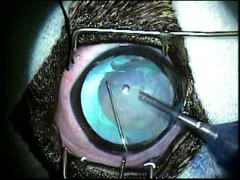
As an opacity of the lens, a cataract can interfere with vision depending on its location and size. When a cataract significantly impairs vision, it can be surgically removed by phacoemulsification.
This is the newest breakthrough in cataract surgery in people and dogs, and is the most common surgical procedure we do.
Phacoemulsification is a specialized, state-of-the-art surgery which utilizes the same equipment, surgical instruments, and medications used on people undergoing cataract surgery.
Cataract phacocmulsification uses ultrasound to break (emulsify) the cataract into very small particles that are aspirated out of the eye. The lens material is carefully and completely sculpted from its capsular bag.
A laser, which is a beam of light, cannot remove a cataract. The entire surgery is performed through a very small corneal incision (3.2mm) at the top of the eye with the aid of an operating microscope and specialized ophthalmic instruments.
The small incision and lens phacoemulsification allows shortened surgical time, less tissue manipulation, and less tissue trauma. This provides for a more rapid physical rehabilitation and improves success.
Another major advantage is that cataract phacoemulsification causes little to no pain. In fact, some dogs with cataract-induced inflammation appear to be more comfortable after surgical recovery.
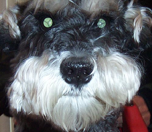
The cataract will not grow back. However, a thin membrane (the posterior lens capsule) that is left intact at the time of surgery may become cloudy. This does not usually appear to interfere with the dog's vision. In some patients, there are capsular opacities and capsular wrinkles even before surgery.
One or both eyes can have cataract surgery at the same time, and some patients are over 15 years of age. A complete ophthalmic and physical examination before surgery is essential.
Presurgical blood and urine tests are also required.
Occasional patients may also need a presurgical retinal test called an electroretinogram if there is concern about retinal function or an ocular ultrasound if there is concern about a retinal detachment.
The usual stay in the hospital is 4 days, but the stay can be as short as 1 night or as long as 2 weeks. The advantage of longer hospitalizations is that we can monitor your pet extremely carefully for any postoperative complications which can significantly improve the surgical outcome.
Eyedrops are needed 6 times a day for the first postoperative month. Medications are then slowly tapered to a low maintenance dose.
Working owners are able to provide these treatments.
Patients begin to see immediately after surgery if there are no complications. Vision continues to improve over the next 6 weeks.
Postoperative examinations are scheduled approximately 1 week, 1 month, and 2 to 3 months after discharge, and then twice yearly thereafter. Follow-up evaluations are extremely important.
The success rate with phacoemulsification is very high. While it is impossible to guarantee the outcome in any particular case, at Animal Eye Care Clinic the success rate for vision is 80 to 93%.
When surgery is successful, your pet's behavior will return to normal or almost normal because of the dramatic improvement in vision, especially if both eyes had cataracts.
However, vision may not be perfect unless an artificial lens implant is placed. The surgical success rate is highest when the cataract is removed before it causes any intraocular damage. Presurgical inflammation and subluxated cataracts may increase the incidence of postoperative complications. The most common temporary postoperative complications are increased intraocular pressure and poor pupil dilation.
Possible blinding complications include severe intraocular inflammation or bleeding, a severe intraocular infection, corneal ulceration, retinal detachment, glaucoma, and intraoperative complete lens luxation. Retinal detachments and glaucoma can occur months to years after surgery. Some of these complications can result in loss of the eye. Occasionally, a small region of the iris may adhere to the lens capsule. This may alter the pupil shape, but it does not interfere with vision.
If a cataract is not removed, it can lead to inflammation, lens dislocations, glaucoma, and retinal detachment with bleeding. Many of these problems will cause your pet pain and irreversible blindness. Therefore, nonsurgical cataracts require monitoring and care.
Remember that if you elect not to do surgery on your pet's cataracts, they need to be monitored since they can lead to inflammation and infection of the eye. Another good reason for at least yearly physicals.
Lens Implants: (Cataract Replacement with a new lens)
By Dr Pentlarge
Intraocular lens (IOL) implants are used after cataract surgery to replace the removed cataractous lens. The purpose of IOL implants is to correct postoperative aphakic (without a lens) vision. In human patients, it is routine to place an IOL implant as part of their cataract surgery unless there is a contraindication. Intraocular lens implants are another new breakthrough in improving the surgical results of cataract surgery.
Patients without a lens are farsighted and may lack crisp vision.
However, canine patients that do not have an IOL implant still demonstrate a dramatic improvement in their visual behavior after successful cataract surgery. This dramatic improvement is especially obvious in patients that were completely blind before surgery.
However, vision will not be perfect or close to perfect without a lens implant. Yet some patients without lens implants will behaviorally act like they have normal vision or you may notice that your pet visually struggles in some situations.
The general consensus among the investigators who developed and are now using IOL implants in dogs is that the IOL implant does improve the dog's vision. This makes sense if we extrapolate from human patients. However, we cannot use human IOL implants because the dog's lens is much larger and has a much higher optical power than the human lens.
Therefore, we use specially developed IOL implants for the dog that are of correct size, are of high quality, and that correct canine aphakic vision. Animal Eye Care Clinic has been placing lens implants in the majority of our cataract patients for the past several years.
The IOL is inserted into the capsular bag after removal of the entire cataractous lens by phacoemulsification-ilTigation-aspiration. The corneal incision is enlarged to 8mm for insertion of the IOL. A viscoelastic material especially designed for cataract surgery is used to protect the corneal endothelium during insertion of the IOL.
Displacement of an IOL implant at some time after surgery has been reported in both human and canine patients, especially if ocular trauma occurs.
Besides improving postoperative vision, clinical evidence shows that the IOL implants may decrease the amount of opacification of the posterior lens capsule. This would further improve vision.
As investigations continue, we will learn more. In the future, we may even have injectable liquid or foldable IOL implants for the dog. Please do not hesitate to ask us any questions or to discuss with us any concerns. There is an additional fee for the intraocular lens implants. We are very excited about this new breakthrough in canine cataract surgery!
Home How we treat different medical problems in pets; What to Expect FoxNest Hospital About our No Kill Shelter
There is a complete site map at the bottom of this page
 | ||||||
Website Directory
Home The Human-Animal Bond The History of Veterinary Medicine About our No Kill Shelter The FoxNest Veterinary Hospital
"What To Expect When You Go To The Vet"
if your pet should have a problem with ...
To include Femoral Head Removal, Hip Dysplasia, Anterior Cruciate Ligament Injuries, Panosteitis, Radiographic Demonstrations, Disc Disease, and Bone Surgery
Strokes, Vascular Diseases, Anemias, DVT, DIC, Blood Parasites, Rat Poison, & Bleeding disorders
Cardiology Heart disease in Cats, Cardiac Hypertrophy, Valvular disease, Cardiac Insufficiency, Congestive Heart Failure, Heartworm Disease, and a little history about the milestones in treating heart disease
Cats: general information page and directory of diseases and problems specific to cats including vaccine recommendations, leukemia, feline viral infections, feline upper respiratory disease and cats that just aren't feeling well.
Dermatology: Skin problems including allergies, rashes, bacterial infections, and itching. Hair Loss, Yeast Infections, Hormonal Problems
Heart disease; Cardiac diseases, vascular diseases, stroke, & heartworms
Hormone Diseases: Diabetes, Thyroid Disease, Cushing's Disease or Hypercortisolism, Addison's disease or Hypocortisolism, Pancreatitis, obesity as a disease
Infectious Diseases Colds, Distemper, Parvo, Leptospirosis, Bruceellosis, Panleukopenia, Feline AIDS, Leukemia, Hepatitis, Kennel Cough, Ringworm, Rabies, FIP, Canine Herpes, Toxic Shock Syndrome, & More
Intestinal problems: diarrhea, constipation, torsion, indigestion, and gas. Also pancreatitis, vomiting, esophagitis, colitis, parvo and other types of dysentery
Metabolic Diseases: Diabetes, Thyroid Disease, Cushing's Disease or Hypercortisolism, Addison's disease or Hypocortisolism, Pancreatitis, obesity as a disease
Neural Problems and Diseases: Epilepsy, Rabies, Distemper, FIP, Paralysis, Tetanus, Seizures, Disc Disease, Toxoplasmosis & others
Parasite Problems Fleas, Ticks, Heartworms, Intestinal Worms, Mosquitos, Lice, Mites, and other welfare recipients
Poisons Snakes, Insects, household chemicals, plants, and foods that might poison your pet
Skeletal-Muscular Problems Arthritis, Fractures, ACL, Ligament Injuries, Disc Disease, Pannus, and many other problems of the bones, muscles, tendons, and ligaments
Skin Problems: allergies, rashes, bacterial infections, and itching. Hair Loss, Yeast Infections, Hormonal Problems
Surgery: Spays, Castrations, Testicle Recipes, Soft Tissue Surgery, Hard Tissue Surgery (Bones), C- Sections, Declawing, Tumor Removal and Cancer Surgery
Other Topics on This Site
Zoonotics: Diseases, worms, and parasites people get from pets.
Includes information about Prescription diets used to treat disease, and a discussion about the pet food industry
Includes information about feline and canine heat or estrus, breeding, C-Sections, pyometra or Infected Uterus, dystocia, no milk, mastitis, & brucellosis
Also newborn care, undescended testicles, and alternative to spaying and castration
WildLife Page: Taking care of baby bunnies, squirrels, and birds. A very funny story about beavers, and other misc information
Our Dog Page: a directory of problems of concern in dogs including parvovirus, distemper, canine herpes, and other diseases